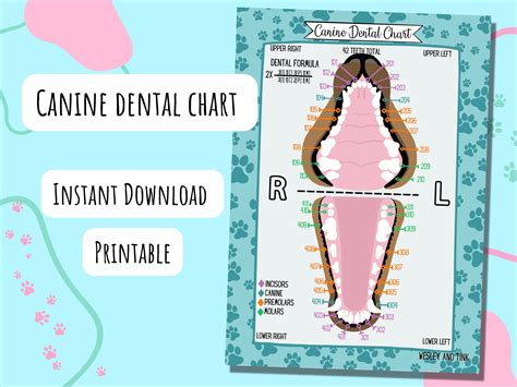
Oh, the joy of a happy, healthy dog! That wagging tail, those soulful eyes, the way they greet you at the door... it’s pure, unconditional love. But beneath that adorable exterior, there's often a silent battle brewing that many of us, as loving pet parents, might overlook: their dental health. Trust me, it’s a game-changer. I remember the first time my vet mentioned my beloved Labrador, Buster, needed a dental cleaning. I felt a pang of guilt, wondering what I had missed. Then, they showed me a printable canine dental chart, and suddenly, the complex world inside his mouth clicked into place. This wasn't just about bad breath; it was about preventing pain, infection, and even serious systemic diseases.
This comprehensive guide is born from countless hours of research, candid conversations with veterinary professionals, and my own journey navigating the ups and downs of pet dental care. My goal is to empower you, whether you’re a first-time dog owner, a seasoned vet tech, or a curious veterinary student, to truly understand, utilize, and even customize a printable canine dental chart. We’re going to demystify canine oral anatomy, equip you with the knowledge to spot common issues, and show you how this simple, yet powerful, tool can be your best friend in advocating for your dog’s long-term health and happiness. Get ready to dive deep into the fascinating world of canine dentistry – your dog's future smile depends on it!
---
Table of Contents

- [The "Why": Unpacking the Critical Importance of Canine Dental Health](#the-why-unpacking-the-critical-importance-of-canine-dental-health)
- [Decoding the Chart: A Tooth-by-Tooth Guide to Canine Dental Anatomy](#decoding-the-chart-a-tooth-by-tooth-guide-to-canine-dental-anatomy)
- [Beyond the Basics: Common Canine Dental Conditions & What to Look For](#beyond-the-basics-common-canine-dental-conditions--what-to-look-for)
- [Charting Like a Pro: Symbols, Notations, and the Art of Accurate Recording](#charting-like-a-pro-symbols-notations-and-the-art-of-accurate-recording)
- [Bringing it Home: Using Your Printable Chart for At-Home Monitoring & Early Detection](#bringing-it-home-using-your-printable-chart-for-at-home-monitoring--early-detection)
- [The Digital Edge: Integrating Printable Charts with Modern Veterinary Practices](#the-digital-edge-integrating-printable-charts-with-modern-veterinary-practices)
- [Customizing Your Chart: Tailoring Templates for Specific Needs](#customizing-your-chart-tailoring-templates-for-specific-needs)
- [When to Call the Vet: Red Flags and Professional Intervention](#when-to-call-the-vet-red-flags-and-professional-intervention)
- [Beyond the Chart: A Holistic Approach to Long-Term Canine Dental Wellness](#beyond-the-chart-a-holistic-approach-to-long-term-canine-dental-wellness)
- [How to Choose the Best Printable Canine Dental Chart for Your Needs](#how-to-choose-the-best-printable-canine-dental-chart-for-your-needs)
- [Common Pitfalls to Avoid When Using Canine Dental Charts](#common-pitfalls-to-avoid-when-using-canine-dental-charts)
- [Advanced Tips for Experts: Optimizing Your Use of Canine Dental Charts](#advanced-tips-for-experts-optimizing-your-use-of-canine-dental-charts)
- [Conclusion](#conclusion)
---
The "Why": Unpacking the Critical Importance of Canine Dental Health

Let's face it, nobody likes bad breath, especially not when it's coming from your beloved canine companion. But beyond the unpleasant odor (halitosis, if we're getting technical!), poor dental health in dogs is a gateway to a myriad of serious, often painful, and sometimes life-threatening issues. Understanding this "why" is the first, crucial step in appreciating the power of a printable canine dental chart.
1. Preventing Pain and Discomfort: Imagine having a constant toothache, or gums that bleed every time you eat. This is the reality for countless dogs suffering from untreated dental disease. They can’t tell us in words, but their reluctance to eat, pawing at their mouth, or simply being less playful are often subtle cries for help.
2. Stopping Systemic Infections: The mouth is not an isolated system. Bacteria from infected gums and teeth can enter the bloodstream and travel to vital organs like the heart, kidneys, and liver, causing severe, sometimes irreversible damage. My own vet once told me about a seemingly healthy dog who developed kidney disease primarily due to chronic dental infections.
3. Avoiding Costly and Complex Treatments: Early detection and intervention, often guided by consistent use of a printable canine dental chart, can prevent the progression of dental disease, potentially saving you thousands in advanced surgical procedures, extractions, and specialized treatments down the line. A little prevention goes a long way!
4. Enhancing Quality of Life: A dog with a healthy mouth can eat comfortably, play with chew toys, and generally enjoy life without the constant nagging pain of dental disease. It’s hard to imagine our furry friends silently suffering from a toothache, isn't it?
5. Understanding Breed Predispositions: Certain breeds, especially smaller ones like Yorkshire Terriers, Chihuahuas, and Poodles, are notoriously prone to dental issues due to crowded teeth and genetic factors. Knowing this helps tailor preventive care, and a chart becomes even more indispensable.
6. Educating Pet Parents: Many pet owners are simply unaware of the severity of dental disease. A well-maintained printable canine dental chart can be an invaluable tool for veterinarians to visually explain their dog's condition, making it much easier for owners to understand and commit to necessary treatments.
7. Establishing a Baseline: By regularly using a chart, you establish a baseline of your dog’s oral health. This makes it easier to spot changes, no matter how subtle, and address them promptly.
8. Monitoring Treatment Progress: After a dental cleaning or extraction, the chart becomes a vital tool for monitoring healing and ensuring the issue doesn’t recur. It’s like having a progress report for their mouth!
9. Improving Overall Health & Longevity: When you manage dental health proactively, you contribute significantly to your dog's overall well-being, energy levels, and ultimately, their lifespan. It's truly a holistic approach.
10. Facilitating Communication: Whether you're a pet owner talking to your vet, or a vet tech communicating with the vet, a standardized dental chart ensures everyone is on the same page, speaking the same "dental language."
11. Recognizing Silent Symptoms: Dogs are masters at hiding pain. A diligent check, even a quick at-home glance guided by a chart, can reveal issues before they become unbearable for your dog.
12. The "Snuggle Factor": Let's be honest, a dog with fresh breath is a much more pleasant snuggling companion! It might sound trivial, but it enhances the bond. My dog, Luna, once had such bad breath I dreaded her kisses. After her dental, it was like having a new dog – kissable and happy!
Decoding the Chart: A Tooth-by-Tooth Guide to Canine Dental Anatomy
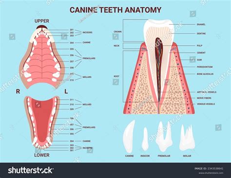
To effectively use a printable canine dental chart, you need to understand what those little squares and lines represent. It’s like trying to explain a complex medical issue without a map – that’s what it’s like without understanding the dental chart! Canine dental anatomy is fascinating, and once you grasp the basics, the chart becomes an intuitive tool. Dogs, like humans, have different types of teeth, each serving a specific purpose.
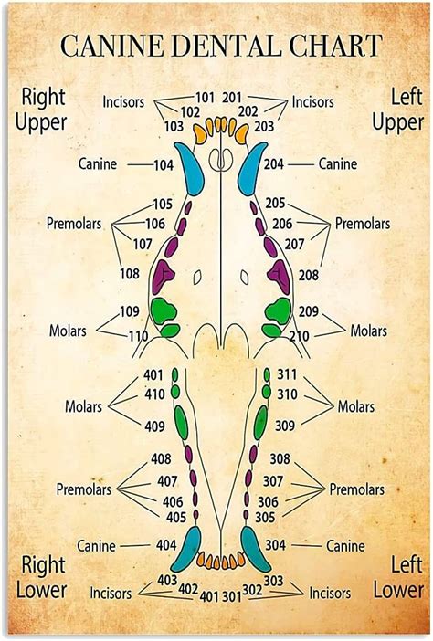
1. The Triadan System: Your Dental GPS: This is the universal numbering system used in veterinary dentistry. It assigns a three-digit number to each tooth. The first digit indicates the quadrant of the mouth (1 for upper right, 2 for upper left, 3 for lower left, 4 for lower right in adults; 5-8 for deciduous/puppy teeth). The last two digits identify the specific tooth. For example, 104 is the upper right canine tooth, and 309 is the lower left first molar. This system ensures clarity and consistency across all veterinary practices.
2. Incisors (I): The Nibblers: Located at the very front of the mouth, dogs typically have 6 upper and 6 lower incisors. These small teeth are primarily used for nibbling, grooming, and picking up small objects. On a chart, they are usually numbered 01, 02, 03 for each quadrant (e.g., 101, 102, 103 for the upper right).
3. Canines (C): The Puncturing Powerhouses: These are the long, pointy teeth on either side of the incisors, giving dogs their characteristic "fang" appearance. There's one upper and one lower canine in each quadrant (four total). They are essential for grasping, tearing, and holding prey. Their deep roots make them very strong. You'll find them as 04 on the chart (e.g., 104, 204, 304, 404).
4. Premolars (P): The Grinding Grinders (Front): Located behind the canines, dogs have four upper and four lower premolars in each quadrant (a total of 16). These teeth have multiple cusps (points) and are used for shearing and grinding food. They are numbered 05, 06, 07, 08. The upper fourth premolar (108, 208) and lower first molar (309, 409) are often called the "carnassial teeth" – the largest and most powerful teeth in a dog's mouth, crucial for crushing bones and tough foods.
5. Molars (M): The Grinding Grinders (Back): These are the flat, broad teeth at the very back of the mouth, designed for crushing and grinding. Dogs typically have two upper molars and three lower molars in each quadrant (total of 10-12, depending on breed). They are numbered 09, 10, 11 (if present).
6. Total Tooth Count: An adult dog typically has 42 permanent teeth (20 upper, 22 lower). Puppies, on the other hand, have 28 deciduous (milk) teeth. A good printable canine dental chart will often have sections for both.
7. Deciduous Teeth (Puppy Teeth): These are temporary teeth that erupt at around 3-4 weeks of age and are replaced by permanent teeth between 3-7 months. Puppy charts use the Triadan system but with the first digit starting at 5 for the upper right, 6 for upper left, 7 for lower left, and 8 for lower right (e.g., 504 is the upper right puppy canine). Retained deciduous teeth are a common issue and can lead to malocclusion and periodontal disease.
8. Root Structure: It's not just the crown you see! Many teeth have multiple roots (e.g., molars can have 2 or 3 roots), which is important for understanding complex extractions and dental X-rays. A chart might indicate root numbers.
9. Gingiva (Gums): The gums are crucial. Healthy gums are pink and firm. Redness, swelling, or bleeding are signs of gingivitis or periodontal disease. Charts often have sections to note gum health around each tooth.
10. Periodontal Ligament: This fibrous tissue connects the tooth root to the bone. Damage to this ligament is a hallmark of periodontal disease.
11. Alveolar Bone: The bone that supports the teeth. Bone loss around the teeth is a serious sign of advanced periodontal disease.
12. Oral Mucosa: The lining of the rest of the mouth, which should also be checked for lesions or masses.
Understanding these components turns a simple printable canine dental chart into a detailed map of your dog's oral health, allowing you to identify specific teeth, note their condition, and track changes over time.
Beyond the Basics: Common Canine Dental Conditions & What to Look For
Knowing the anatomy is one thing; understanding the problems that can arise is another. A printable canine dental chart serves as an indispensable tool for documenting and visualizing these common conditions, helping you and your vet track the progression and treatment.
1. Plaque and Tartar (Calculus) Accumulation:
- What it is: Plaque is a sticky film of bacteria that constantly forms on teeth. If not removed, it hardens into tartar (calculus), a rough, yellowish-brown mineral deposit.
- What to look for: Yellow or brown buildup on the tooth surface, especially near the gum line.
- Charting: Often noted with symbols like "C" or "CAL" and a severity score (1-4).
2. Gingivitis:
- What it is: The earliest stage of periodontal disease, characterized by inflammation of the gums caused by plaque accumulation. It’s reversible with proper cleaning.
- What to look for: Red, swollen, or bleeding gums, especially along the gum line where the tooth meets the gum.
- Charting: Gums are assessed for redness (erythema), swelling (edema), and bleeding on probing (BOP). My own vet noted my Golden Retriever, Finn, had mild gingivitis on his printable canine dental chart, prompting me to start daily brushing.
3. Periodontal Disease:
- What it is: The progression of gingivitis to involve the deeper supporting structures of the tooth, including the periodontal ligament and alveolar bone. This stage is irreversible and can lead to tooth loss.
- What to look for: Receding gums, loose teeth, chronic bad breath, pus around the tooth, pain when chewing.
- Charting: Involves measuring pocket depths (PD) around each tooth, noting bone loss (BL), and tooth mobility (M). It’s heartbreaking, but I once saw a dog with such severe gingivitis, his gums bled just from a gentle touch. The chart helped us track progress after his extensive dental cleaning.
4. Fractured Teeth:
- What it is: Broken teeth, often due to chewing on hard objects (bones, antlers, ice cubes). Fractures can expose the sensitive pulp, leading to pain and infection.
- What to look for: Visible crack or missing piece of a tooth, discoloration (pink/purple if pulp is exposed), reluctance to chew.
- Charting: Noted with "Fx" or "F" and a description (e.g., Fx-crown, Fx-root).
5. Retained Deciduous (Puppy) Teeth:
- What it is: When a puppy tooth doesn't fall out as the permanent tooth erupts, leading to two teeth occupying the same space. This traps food and predisposes to periodontal disease and malocclusion.
- What to look for: Two teeth (a puppy tooth and a permanent tooth) in the same spot.
- Charting: Indicated with "RD" and the tooth number (e.g., RD 504 if the puppy canine is retained).
6. Oral Masses/Tumors:
- What it is: Any abnormal growth in the mouth – can be benign (e.g., epulides) or malignant (e.g., melanoma, squamous cell carcinoma).
- What to look for: Lumps, bumps, discolored areas, or ulcers on the gums, tongue, or inside of the cheeks.
- Charting: Noted with "Mass" or "Lesion" and its location, size, and characteristics.
7. Tooth Abscesses:
- What it is: A pocket of pus caused by a bacterial infection, often at the root tip of a diseased or fractured tooth.
- What to look for: Swelling on the face (often below the eye for an upper carnassial tooth), pain, fever, lethargy.
- Charting: Indicated with "Abs" or "ABSC" on the affected tooth.
8. Attrition and Abrasion:
- What it is: Attrition is tooth wear from tooth-on-tooth contact (e.g., malocclusion). Abrasion is wear from external objects (e.g., chewing on cages, rocks).
- What to look for: Flattened or worn-down tooth surfaces, sometimes exposing the darker dentin.
- Charting: Noted with "ATT" or "ABR."
9. Resorptive Lesions:
- What it is: A painful condition where tooth structure is progressively destroyed, often starting at the gumline. Common in cats but can occur in dogs.
- What to look for: Pink spots on the tooth, sensitivity, or visible tooth erosion.
- Charting: Noted as "RL" or "FORL" for feline odontoclastic resorptive lesions (sometimes adapted for dogs).
10. Malocclusion (Misalignment):
- What it is: Improper alignment of teeth or jaws. Can lead to trauma (e.g., canine teeth hitting the palate), excessive wear, and predispose to periodontal disease.
- What to look for: Crooked teeth, overbites, underbites, or teeth that don't meet properly.
- Charting: Often noted in a specific section for occlusion or with arrows indicating abnormal contact.
11. Oral Foreign Bodies:
- What it is: Objects lodged in the mouth or between teeth (e.g., sticks, bone fragments).
- What to look for: Pawing at the mouth, drooling, reluctance to eat, visible object.
- Charting: Location and type of foreign body.
12. Hypodontia/Oligodontia (Missing Teeth):
- What it is: Congenital absence of one or more teeth.
- What to look for: Gaps where teeth should be.
- Charting: Noted with "M" or "Missing" on the tooth number.
By familiarizing yourself with these conditions and how they are typically represented on a printable canine dental chart, you’ll be much better equipped to understand your vet’s findings and participate actively in your dog’s dental care journey.
Charting Like a Pro: Symbols, Notations, and the Art of Accurate Recording

A printable canine dental chart isn't just a list of teeth; it's a sophisticated shorthand for documenting complex oral health findings. Learning the common symbols and notations is like learning a secret language that unlocks crucial insights into your dog's mouth. While charts can vary slightly, many core symbols are standardized.
1. Missing Tooth (M): Often indicated by drawing an "X" through the tooth or writing "M" next to it. This denotes a tooth that is congenitally absent or has been extracted.
2. Extracted Tooth (EX): Typically marked with an "EX" or a circle around the tooth number, indicating it was present but has been removed. Sometimes, a line through the tooth on the diagram.
3. Fractured Tooth (Fx or F): A common notation for a broken tooth. Often accompanied by a subscript describing the type of fracture (e.g., Fx-crown for a crown fracture, Fx-root for a root fracture, or complicated vs. uncomplicated).
4. Calculus/Tartar (C or CAL): Usually denoted by "C" or "CAL" with a severity score (1-4, where 1 is mild and 4 is severe) next to the tooth, or by shading the tooth on the diagram.
5. Gingivitis (G): Often indicated by "G" with a severity score (0-3, where 0 is healthy, 3 is severe inflammation) or by coloring the gum line red. You might see "E" for erythema (redness) and "ED" for edema (swelling).
6. Periodontal Pocket Depth (PD): Measured in millimeters (mm) with a periodontal probe. These numbers are written next to the tooth, often at multiple points around the tooth (e.g., 5mm, 3mm, 6mm). Deeper pockets indicate bone loss.
7. Gingival Recession (GR): The measurement in mm from the cementoenamel junction (where the crown meets the root) to the free gingival margin. Indicates gum tissue loss.
8. Mobility (M): Graded on a scale of 0-3, where 0 means no mobility, and 3 means severe mobility (the tooth can be moved significantly). Noted as "M0," "M1," "M2," or "M3."
9. Furcation Involvement (F): Refers to bone loss around the roots of multi-rooted teeth. Graded F1, F2, F3 depending on the extent of bone loss in the furcation area.
10. Pulp Exposure (PE): Indicated when the sensitive inner part of the tooth (pulp) is exposed due to a fracture or wear. Often noted with "PE" and may require a root canal or extraction.
11. Caries/Cavity (CA): While less common in dogs than humans, cavities can occur. Noted with "CA" on the tooth.
12. Resorptive Lesions (RL): Marked with "RL" and a type (e.g., Type 1, Type 2) if identified, indicating painful tooth destruction.
13. Occlusion (O): A general section or specific notations to describe how the upper and lower teeth meet. Malocclusions are important to note as they can cause trauma or abnormal wear.
14. Oral Lesions/Masses: Often sketched directly onto the chart's diagram of the mouth, with a description of size, color, and texture in the notes section.
15. Notes Section: This is crucial! It allows for free-form descriptions of unusual findings, client concerns, treatment plans, and follow-up recommendations. This is where the narrative of the oral exam unfolds. For my dog, Buster, his chart had extensive notes detailing the extent of his periodontal disease and the specific extractions performed.
16. Date and Examiner: Always include the date of the exam and the initials or name of the person performing the charting. This ensures proper record-keeping and accountability.
Mastering these symbols transforms a blank printable canine dental chart into a dynamic diagnostic tool, allowing for precise communication between veterinary staff and clear understanding for pet owners.
Bringing it Home: Using Your Printable Chart for At-Home Monitoring & Early Detection
You don't need to be a vet to use a printable canine dental chart effectively at home. In fact, for pet owners, it’s one of the most proactive steps you can take for your dog's health between professional cleanings. This simplified approach focuses on observation and early detection.
1. Establish a Routine: Choose a consistent time each week or month for a quick oral check. Maybe during cuddle time, or after a walk. Consistency is key to noticing subtle changes.
2. Gather Your Tools: All you really need is your printable canine dental chart, a pen, and good lighting. A headlamp or flashlight can be surprisingly helpful.
3. Positive Reinforcement: Make it a positive experience for your dog. Start slowly, praise them, and offer treats. The goal is to make them comfortable with their mouth being handled. My neighbor, Sarah, used a simplified chart I shared with her and caught a fractured tooth on her terrier early, saving him a lot of pain and a more complex procedure.
4. Check for Bad Breath (Halitosis): While not a charting symbol, persistent, foul breath is often the first sign of dental disease. Note it down.
5. Examine the Gums: Gently lift your dog’s lips to expose their gums. Healthy gums are pink and firm, fitting snugly around the teeth.
- What to chart: Note any redness, swelling, or bleeding. If you see bright red or purple gums, mark it on the chart and consider it a red flag.
6. Inspect Each Tooth for Tartar: Look for yellow, brown, or green-brown buildup, especially on the back teeth and near the gum line.
- What to chart: Use a simple shading system or a "C" for calculus on the corresponding tooth on your printable canine dental chart. A scale of 1-3 can be helpful (1=mild, 3=heavy).
7. Look for Broken or Chipped Teeth: Carefully examine all visible teeth for any chips, cracks, or missing pieces. Also, check for any discolored teeth (pink, purple, or grey), which can indicate a dead pulp.
- What to chart: Draw a simple "X" for a missing tooth, or a line through a broken tooth, noting "chip" or "Fx" in your notes.
8. Check for Loose Teeth: Gently (and carefully!) try to wiggle each tooth. Any noticeable movement is a concern.
- What to chart: Mark "M" for mobility on the affected tooth.
9. Scan for Oral Masses or Lesions: Look at the inside of the cheeks, the roof of the mouth, and under the tongue for any unusual lumps, bumps, sores, or discolored patches.
- What to chart: Make a note of the location and description on your printable canine dental chart or on the general mouth diagram.
10. Note Any Changes in Behavior: Is your dog suddenly reluctant to chew, dropping food, favoring one side of their mouth, or pawing at their face? These are indirect signs of pain.
- What to chart: Add a note in a general comments section.
11. Document Your Findings: Don't just observe; write it down! Even if it's just "slight redness on 208 gum," these notes over time create a valuable history.
12. Share with Your Vet: Bring your filled-out printable canine dental chart to your dog's annual check-up. It provides your vet with invaluable insights and shows your commitment to your dog’s health, making their job easier and your dog's care more precise. This proactive approach allows for early detection, which can often mean less invasive and less costly treatments down the line.
The Digital Edge: Integrating Printable Charts with Modern Veterinary Practices

While the tangible nature of a printable canine dental chart is incredibly valuable, especially for hands-on learning and quick reference, modern veterinary practices are increasingly leveraging digital solutions. However, the printable format still plays a crucial role, often serving as a bridge or a foundational component for these advancements.
1. Digital Charting Software Integration: Many veterinary practice management software systems now include integrated dental charting modules. These digital charts are based on the same principles as the printable ones, using the Triadan system and standard symbols, but offer enhanced features.
2. The "Print-to-Digital" Workflow: Often, a blank printable canine dental chart is used during the initial hands-on examination in the operating room. Findings are quickly jotted down, and then transcribed into the digital system later. This allows the vet or tech to focus on the patient without fumbling with a computer.
3. Enhanced Data Storage and Retrieval: Digital charts allow for easy storage, searchability, and retrieval of historical dental records. Imagine trying to find a specific paper chart from five years ago in a busy clinic! This greatly streamlines patient care and follow-up.
4. Improved Legibility and Consistency: No more deciphering messy handwriting! Digital charting ensures all entries are clear and standardized, reducing errors and misinterpretations.
5. Integration with Electronic Medical Records (EMR): Digital dental charts are seamlessly integrated with a patient’s overall EMR, providing a holistic view of their health. This allows for cross-referencing with blood work, medication history, and other diagnostic